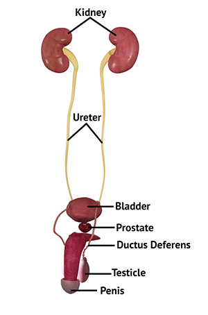
Anatomy of the Male Urinary System
The male urinary system consists of several organs including the kidneys, ureters, bladder, and urethra, which work together to produce and assist in the release of waste. Two bean-shaped kidneys filter the blood of waste products to form urine. Each kidney is differentiated into the outer cortex and inner medulla and is filled with special units called nephrons. Urine accumulates at the funnel-shaped opening of the kidney called the renal pelvis. The renal artery, vein, and nerves enter the kidneys at the hilum. The renal pelvis at each kidney opens out into a long tube called a ureter. The ureters are lined with smooth muscle and carry urine to the bladder through openings called ureteral orifices in the bladder wall.
The bladder is present within the pelvis and shaped like a pyramid. It is lined by smooth muscles and can expand to store about 500 mL of urine. Urine is stored until the bladder gets full. This signals the brain, which in turn sends signals to ring-like muscles called the internal sphincter muscles lining the opening of the bladder to relax and allow the outflow of urine through the urethra. The urethra is a tube through which urine passes out from the bladder, and is longer in males. Its initial segment passes through the prostate gland and contains openings for sperm and fluids that form semen. The next segment of the urethra is surrounded by an external sphincter that helps provide further urine retention. The final segment of the urethra runs through the penis and opens out through the urethral opening at the tip of the penis.
The urinary system is designed in such a way that it only allows the one-way movement of urine from the kidneys to the outside. Abnormalities and blockages in any part of the urinary system may enable the urine to backflow, causing serious health issues.